Shop
Displaying 1–48 of 467 results
AAAAI Guidelines Bundle
Last Updated: Feb 16, 2023
AAN Guidelines Bundle
Last Updated: Mar 21, 2022
$
54.95
Print
Included Guidelines:
- Antiseizure Medication Withdrawal
- Early Parkinson Disease
- Painful Diabetic Polyneuropathy
- Patent Foramen Ovale
- Prevention, Diagnosis and Treatment of Lyme Disease
- Stroke Prevention in Symptomatic Large Artery Intracranial Atherosclerosis
- The Care of the Patient with Amyotrophic Lateral Sclerosis: Drug, Nutritional, And Respiratory Therapies

AAO-HNS Guidelines Bundle
Last Updated: Feb 27, 2024
$
158.95
Print
Included Guidelines:
- Acute Otitis Externa
- Adult Sinusitis
- Allergic Rhinitis
- Bell’s Palsy
- Benign Paroxysmal Positional Vertigo
- Cerumen Impaction
- Evaluation of the Neck Mass in Adults
- Hoarseness (Dysphonia)
- Immunotherapy for Inhalant Allergy
- Improving Nasal Form and Function after Rhinoplasty
- Improving Voice Outcomes After Thyroid Surgery
- Ménière’s Disease
- Nosebleed (Epistaxis)
- Opioid Prescribing for Analgesia After Common Otolaryngology Operations
- Otitis Media with Effusion
- Sudden Hearing Loss
- Tinnitus
- Tonsillectomy in Children
- Tympanostomy Tubes in Children
ACAAI Guidelines Bundle
Last Updated: Feb 16, 2023
ACC Guidelines Bundle
Last Updated: Nov 30, 2023
$
323.95
Print
Included Guidelines:
- Coronary Artery Revascularization
- Device-Based Therapy
- Diagnosis and Management of Aortic Disease
- Diagnosis and Treatment of Patients with Hypertrophic Cardiomyopathy
- Evaluation and Diagnosis of Chest Pain
- Evaluation and Management of Patients with Bradycardia and Cardiac Conduction Delay
- Evaluation and Management of Patients With Syncope
- Heart Failure
- Management of Adults with Congenital Heart Disease
- Management of Blood Cholesterol
- Management of Patients with Atrial Fibrillation
- Management of Patients with Chronic Coronary Disease
- Management of Patients With Ventricular Arrythmias and the Prevention of Sudden Cardiac Death
- Non-ST-Elevation Acute Coronary Syndromes
- Perioperative Cardiovascular Evaluation and Management of Patients Undergoing Noncardiac Surgery
- Peripheral Artery Disease Lower Extremity
- Prevention, Detection, Evaluation, and Management of High Blood Pressure in Adults
- Primary Prevention of Cardiovascular Disease
- ST-Elevation Myocardial Infarction (STEMI)
- Stable Ischemic Heart Disease
- Supraventricular Tachycardia
- Valvular Heart Disease
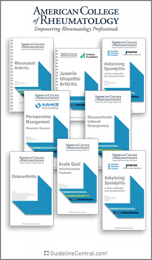
ACR Guidelines Bundle
Last Updated: Dec 19, 2023
$
158.95
Print
Included Guidelines:
- ANCA-Associated Vasculitis
- Ankylosing Spondylitis
- Glucocorticoid‐Induced Osteoporosis
- Juvenile Idiopathic Arthritis
- Juvenile Idiopathic Arthritis Uveitis
- Juvenile Idiopathic Arthritis — Non-Pharmacologic Therapies, Medication Monitoring, Immunizations, and Imaging
- Juvenile Idiopathic Arthritis — Therapeutic Approaches for Oligoarthritis, Temporomandibular Joint Arthritis and Systemic JIA
- Kawasaki Disease
- Lupus Nephritis
- Management of Gout
- Management of Reproductive Health in Rheumatic and Musculoskeletal Diseases
- Osteoarthritis
- Perioperative Management Rheumatic Diseases
- Polyarteritis Nodosa
- Prevention, Diagnosis and Treatment of Lyme Disease
- Psoriatic Arthritis Treatment
- Rheumatoid Arthritis
- Rheumatoid Arthritis Integrative Interventions
- Total Joint Arthroplasty (TJA) of the Hip or Knee
- Vaccinations in Patients with Rheumatic and Musculoskeletal Diseases

AGA Guidelines Bundle
Last Updated: Aug 18, 2023
$
225.95
Print
Included Guidelines:
- Acute Diverticulitis
- Acute Liver Failure
- Acute Pancreatitis
- Adult Obesity — Pharmacological Interventions
- Asymptomatic Neoplastic Pancreatic Cysts
- Biomarkers in Ulcerative Colitis
- Bowel Cleansing
- Chronic Idiopathic Constipation
- Coagulation Disorders in Patients With Cirrhosis
- Colorectal Cancer Screening
- Constipation
- Elastography in the Evaluation of Liver Fibrosis
- Endoscopic Removal of Colorectal Lesions
- Gastric Intestinal Metaplasia
- Gastrointestinal Evaluation of Iron Deficiency Anemia
- Inflammatory Bowel Disease
- Irritable Bowel Syndrome With Constipation
- Irritable Bowel Syndrome With Diarrhea
- Laboratory Evaluation of Functional Diarrhea and Diarrhea-Predominant Irritable Bowel Syndrome in Adults
- Management of Crohn's Disease
- Management of Eosinophilic Esophagitis
- Microscopic Colitis
- Mild-Moderate Ulcerative Colitis Management
- Moderate to Severe Luminal and Fistulizing Crohn’s Disease
- Moderate to Severe Ulcerative Colitis
- Opioid-Induced Constipation
- Pharmacological Management of Irritable Bowel Syndrome
- Role of Probiotics in the Management of Gastrointestinal Disorders
- Systemic Therapy for Hepatocellular Carcinoma
- Upper Gastrointestinal Biopsy
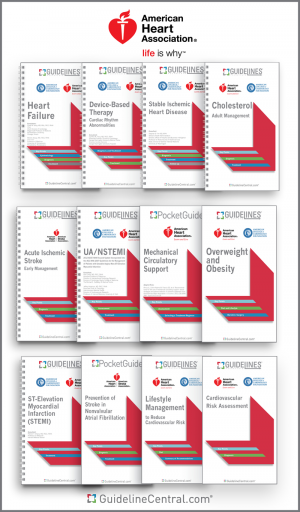
AHA Guidelines Bundle
Last Updated: Nov 30, 2023
$
387.95
Print
Included Guidelines:
- Coronary Artery Revascularization
- CPR for Mechanical Circulatory Support
- Device-Based Therapy
- Diagnosis and Management of Aortic Disease
- Diagnosis and Treatment of Patients with Hypertrophic Cardiomyopathy
- Early Management of Patients With Acute Ischemic Stroke
- Evaluation and Diagnosis of Chest Pain
- Evaluation and Management of Patients with Bradycardia and Cardiac Conduction Delay
- Evaluation and Management of Patients With Syncope
- Heart Failure
- Management of Adults with Congenital Heart Disease
- Management of Blood Cholesterol
- Management of Patients with Atrial Fibrillation
- Management of Patients with Chronic Coronary Disease
- Management of Patients With Ventricular Arrythmias and the Prevention of Sudden Cardiac Death
- Mechanical Circulatory Support
- Mechanical Circulatory Support — Ambulatory and Community Patient Care
- Non-ST-Elevation Acute Coronary Syndromes
- Perioperative Cardiovascular Evaluation and Management of Patients Undergoing Noncardiac Surgery
- Peripheral Artery Disease Lower Extremity
- Prevention Of Stroke In Nonvalvular Atrial Fibrillation
- Prevention of Stroke In Women
- Prevention, Detection, Evaluation, and Management of High Blood Pressure in Adults
- Primary Prevention of Cardiovascular Disease
- Primary Stroke Prevention
- ST-Elevation Myocardial Infarction (STEMI)
- Stable Ischemic Heart Disease
- Supraventricular Tachycardia
- Valvular Heart Disease

AND Guidelines Bundle
Last Updated: Nov 7, 2022
$
80.95
Print
Included Guidelines:
- Adult Weight Management
- Chronic Kidney Disease Evidence-Based Nutrition
- Chronic Obstructive Pulmonary Disease Nutrition Management
- Cystic Fibrosis Evidence-Based Nutrition Recommendations
- Diabetes Medical Nutrition Therapy
- Enteral Nutrition in Preterm Infants
- Gestational Diabetes Mellitus
- Heart Failure
- Hypertension Nutrition Management
- Pediatric Weight Management
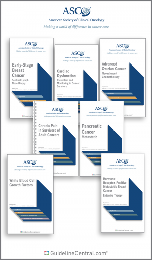
ASCO Guidelines Bundle
Last Updated: Mar 19, 2024
$
774.95
Print
Included Guidelines:
- Adjuvant Bisphosphonates and Other Bone-Modifying Agents in Breast Cancer
- Adjuvant Therapy for Stage III Colon Cancer
- Advanced Gastroesophageal Cancer
- Advanced Human Epidermal Growth Factor Receptor 2-Positive Breast Cancer
- Advanced Human Epidermal Growth Factor Receptor 2-Positive Breast Cancer – Brain Metastases
- Antiemetics
- Antimicrobial Prophylaxis for Adult Patients With Cancer-Related Immunosuppression
- Anxiety and Depression in Adult Survivors of Cancer
- Appropriate Systemic Therapy Dosing for Obese Adult Patients with Cancer
- Axilla Management in Early-Stage Breast Cancer
- Biomarkers for Adjuvant Endocrine and Chemotherapy in Early-Stage Breast Cancer
- Biomarkers for Systemic Therapy in Metastatic Breast Cancer
- Bone-Modifying Agents in Metastatic Breast Cancer
- Bone-Modifying Agents in Multiple Myeloma
- Breast Cancer Germline Testing
- Cancer-Associated Anemia With Erythropoiesis-Stimulating Agents
- Cannabis in Adults with Cancer
- Cardiac Dysfunction - Prevention and Monitoring in Cancer Survivors
- Cervical Cancer Prevention
- Chemotherapy in Combination With Radiotherapy for Definitive-Intent Treatment of Stage II-IVA Nasopharyngeal Carcinoma
- Chemotherapy-Naïve Castration-Resistant Prostate Cancer
- Chronic Pain in Survivors of Adult Cancers
- Clear Cell Renal Cell Carcinoma
- Colorectal Cancer Biomarkers
- Colorectal Cancer Early Detection
- Diagnosis and Management of Squamous Cell Carcinoma of Unknown Primary in the Head and Neck
- Diffuse Astrocytic and Oligodendroglial Tumors in Adults
- Early-Stage Breast Cancer
- Estrogen and Progesterone Receptor Testing in Breast Cancer
- Exercise, Diet, and Weight Management During Cancer Treatment
- Fertility Preservation
- Germline and Somatic Tumor Testing in Epithelial Ovarian Cancer
- Hepatitis B Virus Screening and Management for Patients with Cancer Prior to Therapy
- HER2 Testing in Breast Cancer
- Hormone Receptor–Positive Breast Cancer Adjuvant Endocrine Therapy
- HR-Positive, HER2-Negative Metastatic Breast Cancer
- Immune-related Adverse Events from Immune Checkpoint Inhibitor Therapy
- Immune-related Adverse Events — CAR T-Cell Therapy
- Initial Management of Noncastrate Advanced, Recurrent, or Metastatic Prostate Cancer
- Integrative Medicine for Pain Management in Oncology
- Integrative Oncology Care of Symptoms of Anxiety and Depression in Adult Patients with Cancer
- Invasive Cervical Cancer
- Laryngeal Cancer
- Late-Stage Colorectal Cancer
- Lung Cancer Surveillance After Definitive Curative-Intent Therapy
- Malignant Pleural Mesothelioma
- Management of Cancer Cachexia
- Management of Dyspnea in Advanced Cancer
- Management of Hereditary Breast Cancer
- Management of Male Breast Cancer
- Management of Osteoporosis
- Management of Salivary Gland Malignancy
- Management of the Neck in Squamous Cell Carcinoma of the Oral Cavity and Oropharynx
- Medication-Related Osteonecrosis of the Jaw
- Metastatic Colorectal Cancer
- Metastatic HER2-Negative Breast Cancer – Chemo- and Targeted Therapy
- Metastatic Pancreatic Cancer
- Metastatic Well-differentiated Gastroenteropancreatic Neuroendocrine Tumors
- Molecular Biomarkers in Localized Prostate Cancer
- Multiple Myeloma
- Neoadjuvant Chemotherapy for Newly Diagnosed, Advanced Ovarian Cancer
- Neoadjuvant Chemotherapy, Endocrine Therapy, and Targeted Therapy for Breast Cancer
- Non-Small Cell Lung Cancer — Adjuvant Therapy for Completely Resected Stages I-IIIA
- Optimum Imaging Strategies for Advanced Prostate Cancer
- Outpatient Management of Fever and Neutropenia in Adults Treated for Malignancy
- Ovarian Masses and Treatment of Epithelial Ovarian Cancer
- Palliative Care in the Global Setting
- Pancreatic Cancer - Evaluating Susceptibility
- PARP Inhibitors in the Management of Ovarian Cancer
- Penile Cancer
- Platelet Transfusion
- Potentially Curable Pancreatic Adenocarcinoma
- Prevention And Management Of Chemotherapy-Induced Peripheral Neuropathy In Survivors Of Adult Cancers
- Recurrent and Metastatic Head and Neck Cancers
- Salivary Gland Hypofunction and/or Xerostomia
- Secondary Prevention of Cervical Cancer
- Selection of Optimal Adjuvant Chemotherapy and Targeted Therapy for Early Breast Cancer
- Small Renal Masses
- Somatic Genomic Testing for Metastatic or Advanced Cancer
- Stage II Colon Cancer Adjuvant Therapy
- Stage III Non-Small Cell Lung Cancer
- Stage IV Non-Small Cell Lung Cancer without Driver Alterations
- Systemic Therapy for Advanced Hepatocellular Carcinoma
- Systemic Therapy for Melanoma
- Systemic Therapy for Small Cell Lung Cancer
- Systemic Treatment of Patients With Metastatic Breast Cancer
- Therapy for Stage IV Non-Small-Cell Lung Cancer With Driver Alterations
- Treatment for Brain Metastases
- Treatment of Early-Stage Colorectal Cancer
- Treatment of Locally Advanced Esophageal Carcinoma
- Treatment of Metastatic Carcinoma and Myeloma of the Femur
- Use of Endocrine Therapy for Breast Cancer Risk Reduction
- Use of Opioids for Cancer Pain
- Vaccination of Adults with Cancer
- Venous Thromboembolism Prophylaxis and Treatment in Patients with Cancer
- Vulnerabilities in Older Patients Receiving Systemic Cancer Therapy
- White Blood Cell Growth Factors
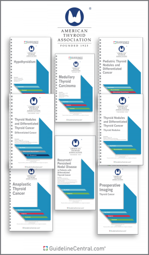
ATA Guidelines Bundle
Last Updated: Apr 4, 2024
$
107.95
Print
Included Guidelines:
- Ablation Techniques for Benign Thyroid Nodules
- Anaplastic Thyroid Cancer
- Hyperthyroidism
- Hypothyroidism
- Medullary Thyroid Carcinoma
- Pediatric Thyroid Nodules and Differentiated Cancer
- Preoperative Imaging Thyroid Cancer
- Recurrent/Persistent Nodal Disease in Patients with Differentiated Thyroid Cancer
- Thyroid Disease During Pregnancy
- Thyroid Hormones
- Thyroid Nodules and Differentiated Thyroid Cancer Differentiated Cancer
- Thyroid Nodules and Differentiated Thyroid Cancer Thyroid Nodules
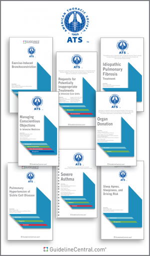
ATS Guidelines Bundle
Last Updated: Feb 2, 2024
$
332.95
Print
Included Guidelines:
- Definition, Evaluation and Treatment of Severe Asthma
- Diagnosis and Detection of Sarcoidosis
- Diagnosis and Treatment of Adults with Community-Acquired Pneumonia
- Diagnosis of Hypersensitivity Pneumonitis in Adults
- Diagnosis of Idiopathic Pulmonary Fibrosis
- Diagnosis of Primary Ciliary Dyskinesia
- Diagnosis, Risk Stratification, And Management Of Pulmonary Hypertension Of Sickle Cell Disease.
- Diagnostic Evaluation of Infants with Recurrent or Persistent Wheezing
- Drug-Susceptible Tuberculosis
- Ethical and Policy Considerations in Organ Donation after Circulatory Determination of Death
- Evaluation and Management of Obesity Hypoventilation Syndrome
- Exercise-induced Bronchoconstriction
- Fractional Exhaled Nitric Oxide for Asthma
- Home Oxygen Therapy for Adults with Chronic Lung Disease
- Home Oxygen Therapy for Children
- Hospital-acquired and Ventilator-Associated Pneumonia
- Idiopathic Pulmonary Fibrosis and Progressive Pulmonary Fibrosis in Adults
- Initiating Pharmacologic Treatment in Tobacco-Dependent Adults
- Liberation from Mechanical Ventilation in Critically Ill Adults
- Long-Term Noninvasive Ventilation in Chronic Stable Hypercapnic Chronic Obstructive Pulmonary Disease
- Management of Adult Patients with Acute Respiratory Distress Syndrome
- Management of Malignant Pleural Effusions
- Managing Conscientious Objections in Intensive Care Medicine
- Mechanical Ventilation in Adult Patients with Acute Respiratory Distress Syndrome
- Microbiological Laboratory Testing in the Diagnosis of Fungal Infections in Pulmonary and Critical Care Practice
- Noninvasive Ventilation For Acute Respiratory Failure
- Nucleic Acid-Based Testing for Non-Influenza Viral Pathogens
- Pediatric Chronic Home Invasive Ventilation
- Persistent, Post-adenotonsillectomy Obstructive Sleep Apnea in Children
- Pharmacologic Management of Chronic Obstructive Pulmonary Disease
- Post-Prematurity Respiratory Disease
- Prevention and Management of COPD Exacerbations
- Pulmonary Rehabilitation for Adults with Chronic Respiratory Disease
- Requests for Potentially Inappropriate Treatments In Intensive Care Units
- Role of Weight Management in the Treatment of Adult Obstructive Sleep Apnea
- Routine Lung Function Tests
- Severe Asthma Management
- Sleep Apnea, Sleepiness, and Driving Risk in Noncommercial Drivers
- Systemic Sclerosis-Associated Interstitial Lung Disease
- Treatment of Drug-Resistant Tuberculosis
- Treatment Of Idiopathic Pulmonary Fibrosis
- Treatment of Nontuberculous Mycobacterial Pulmonary Disease
- Tuberculosis Diagnosis Adults and Children

ES Guidelines Bundle
Last Updated: Feb 9, 2023
$
193.95
Print
Included Guidelines:
- Acromegaly
- Congenital Adrenal Hyperplasia Due to Steroid 21-Hydroxylase Deficiency
- Cushing's Syndrome Treatment
- Diabetes and Pregnancy
- Diagnosis of Cushing's Syndrome
- Evaluation and Treatment of Hypertriglyceridemia
- Functional Hypothalamic Amenorrhea
- Gender-Dysphoria / Gender-Incongruence
- Hirsutism in Premenopausal Women
- Hypercalcemia of Malignancy in Adults
- Hyperglycemia in Hospitalized Adult Patients
- Hypothalamic–Pituitary and Growth Disorders in Survivors of Childhood Cancer
- Lipid Management in Patients with Endocrine Disorders
- Management of Individuals with Diabetes at High Risk for Hypoglycemia
- Menopause
- Obesity
- Paget's Disease of Bone
- Pediatric Obesity
- Pharmacological Management of Osteoporosis in Postmenopausal Women
- Primary Adrenal Insufficiency
- Primary Aldosteronism
- Primary Prevention of ASCVD and T2DM in Patients at Metabolic Risk
- Testosterone Therapy in Men With Hypogonadism
- Treatment of Diabetes in Older Adults
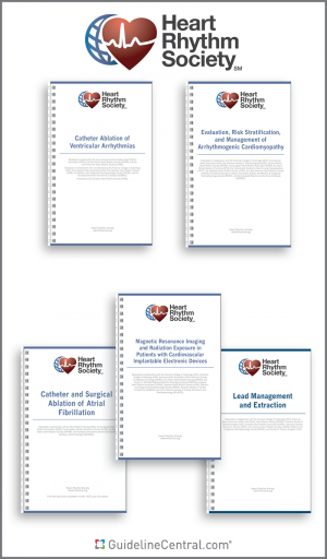
HRS Guidelines Bundle
Last Updated: Nov 30, 2023
$
187.95
Print
Included Guidelines:
- Cardiac Physiologic Pacing for the Avoidance and Mitigation of Heart Failure
- Cardiovascular Implantable Electronic Device Lead Management and Extraction
- Catheter Ablation of Ventricular Arrhythmias
- Catheter and Surgical Ablation of Atrial Fibrillation
- Device-Based Therapy
- Evaluation and Management of Arrhythmic Risk in Neuromuscular Disorders
- Evaluation and Management of Patients with Bradycardia and Cardiac Conduction Delay
- Evaluation and Management of Patients With Syncope
- Evaluation, Risk Stratification, and Management of Arrhythmogenic Cardiomyopathy
- Magnetic Resonance Imaging and Radiation Exposure in Patients with Cardiovascular Implantable Electronic Devices
- Management of Patients with Atrial Fibrillation
- Management of Patients With Ventricular Arrythmias and the Prevention of Sudden Cardiac Death
- Practical Management of the Remote Device Clinic
- Supraventricular Tachycardia
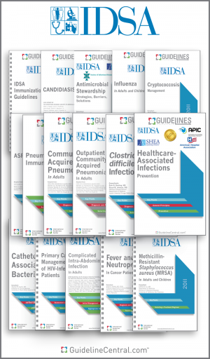
IDSA Guidelines Bundle
Last Updated: Dec 11, 2023
$
408.95
Print
Included Guidelines:
- Acute Hematogenous Osteomyelitis
- Antibiotic Stewardship
- Antimicrobial Prophylaxis for Adult Patients With Cancer-Related Immunosuppression
- Antimicrobial Prophylaxis in Surgery
- Aspergillosis
- Asymptomatic Bacteriuria Management
- Candida
- Catheter-Associated Bacteriuria
- Chronic Kidney Disease in HIV-Infected Patients
- Chronic Pain in HIV
- Clostridium difficile
- Coccidioidomycosis
- Complicated Intra-abdominal Infection in Adults
- Cryptococcosis Management
- Diabetic Foot Infections
- Diagnosis and Treatment of Adults with Community-Acquired Pneumonia
- Drug-Susceptible Tuberculosis
- Fever and Neutropenia in Cancer Patients
- Guideline on Diagnosis and Management of Babesiosis
- Hospital-acquired and Ventilator-Associated Pneumonia
- Infectious Diarrhea
- Influenza Update for the 2020 - 2021 Flu Season
- Leishmaniasis
- Management of Outpatient Parenteral Antimicrobial Therapy
- Methicillin-Resistant Staphylococcus aureus (MRSA)
- Native Vertebral Osteomyelitis in Adults
- Neurocysticercosis
- Outpatient Management of Fever and Neutropenia in Adults Treated for Malignancy
- Pediatric Community-Acquired Pneumonia
- Prevention of Ventilator-Associated Pneumonia, Ventilator-Associated Events, and Nonventilator Hospital-Acquired Pneumonia in Acute Care Hospitals
- Prevention, Diagnosis and Treatment of Lyme Disease
- Primary Care Guidance for Persons with HIV
- Prosthetic Joint Infection
- Rhinosinusitis
- Skin and Soft Tissue Infections
- Strategies to Prevent Catheter-Associated Urinary Tract Infections in Acute-Care Hospitals
- Strategies to Prevent Central Line-Associated Bloodstream Infections in Acute-Care Hospitals
- Strategies to Prevent Clostridioides difficile Infections in Acute-Care Hospitals
- Strategies to Prevent Healthcare-Associated Infections through Hand Hygiene
- Strategies to Prevent Methicillin-Resistant Staphylococcus aureus in Acute-Care Hospitals
- Strategies to Prevent Surgical Site Infections in Acute Care Hospitals
- Streptococcal Pharyngitis Diagnosis and Management
- Treatment of Acute Uncomplicated Cystitis and Pyelonephritis in Women
- Treatment of Drug-Resistant Tuberculosis
- Treatment of Nontuberculous Mycobacterial Pulmonary Disease
- Tuberculosis Diagnosis Adults and Children
- Vaccination of the Immunocompromised Host
- Ventriculitis and Meningitis Healthcare-Associated
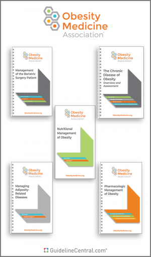
OMA Guidelines Bundle
Last Updated: Apr 11, 2024
PALTC Guidelines Bundle
Last Updated: Dec 19, 2021
$
98.95
Print
Included Guidelines:
- Advance Care Planning (ACP) Discussion Guide And Visit Documentation Template
- COPD Management in the Post-Acute and Long-Term Care Setting
- Delirium, Dementia And Depression In The Long-Term Care Setting
- Diabetes Management Post-Acute and Long-Term Care Setting
- Diagnosis, Treatment, and Prevention of Urinary Tract Infections in Post-Acute and Long-Term Care Settings
- Heart Failure In the Post acute and Long-Term Care Setting
- Pain Management In The Post-Acute And Long-Term Care Setting
- Parkinson’s Disease & Psychosis in the Post-Acute and Long-Term Care Setting
- Pressure Ulcers and other Wounds in the Post-Acute and Long-Term Care Setting
SHEA Guidelines Bundle
Last Updated: Dec 11, 2023
$
159.95
Print
Included Guidelines:
- Animals in Healthcare Facilities
- Antibiotic Stewardship
- Antibiotic Stewardship in Hospitals During Public Health Emergencies
- Antimicrobial Prophylaxis in Surgery
- Clostridium difficile
- Duration of Contact Precautions in Acute Care Settings
- Healthcare Personnel with Hepatitis B, Hepatitis C, or HIV
- Infection Prevention in the Operating Room Anesthesia Work Area
- Isolation Precautions for Visitors
- NICU Clostridioides difficile
- NICU Staphylococcus aureus
- Nursing Home Infections
- Prevention of Central Line-Associated Bloodstream Infections in the Neonatal Intensive Care Unit
- Prevention of Ventilator-Associated Pneumonia, Ventilator-Associated Events, and Nonventilator Hospital-Acquired Pneumonia in Acute Care Hospitals
- Strategies to Prevent Catheter-Associated Urinary Tract Infections in Acute-Care Hospitals
- Strategies to Prevent Central Line-Associated Bloodstream Infections in Acute-Care Hospitals
- Strategies to Prevent Clostridioides difficile Infections in Acute-Care Hospitals
- Strategies to Prevent Healthcare-Associated Infections through Hand Hygiene
- Strategies to Prevent Methicillin-Resistant Staphylococcus aureus in Acute-Care Hospitals
- Strategies to Prevent Surgical Site Infections in Acute Care Hospitals
SVS Guidelines Bundle
Last Updated: Dec 21, 2023
$
110.95
Print
Included Guidelines:
- Abdominal Aortic Aneurysm
- Atherosclerotic Occlusive Disease of the Lower Extremities
- Chronic Limb-Threatening Ischemia
- Chronic Mesenteric Ischemia
- Extracranial Cerebrovascular Disease
- Follow-Up After Vascular Surgery Arterial Procedures
- Lower Extremity Varicose Veins
- Management of Diabetic Foot
- Management of Visceral Aneurysms
- Popliteal Artery Aneurysms
- Reporting Standards for Type B Aortic Dissections
- Thoracic Endovascular Aneurysm Repair
- Venous Leg Ulcers
Abdominal Aortic Aneurysm
Last Updated: Jan 30, 2018
Ablation Techniques for Benign Thyroid Nodules
Last Updated: Jan 24, 2024
Acromegaly
Last Updated: Jan 29, 2015
Acute Diverticulitis
Last Updated: Oct 10, 2016
Acute Hematogenous Osteomyelitis
Last Updated: Dec 1, 2021
Acute Liver Failure
Last Updated: Mar 1, 2017
Acute Otitis Externa
Last Updated: Oct 23, 2014
Acute Pancreatitis
Last Updated: May 7, 2018
Adjuvant Bisphosphonates and Other Bone-Modifying Agents in Breast Cancer
Last Updated: Jan 16, 2022
Adjuvant Therapy for Stage III Colon Cancer
Last Updated: Apr 14, 2019
Adult Obesity — Pharmacological Interventions
Last Updated: Feb 15, 2023
Adult Sinusitis
Last Updated: Apr 1, 2015
Adult Weight Management
Last Updated: Nov 6, 2022
Advance Care Planning (ACP) Discussion Guide And Visit Documentation Template
Last Updated: Mar 25, 2018
Advanced Gastroesophageal Cancer
Last Updated: Jan 5, 2023
Advanced Human Epidermal Growth Factor Receptor 2-Positive Breast Cancer
Last Updated: May 31, 2022
Advanced Human Epidermal Growth Factor Receptor 2-Positive Breast Cancer – Brain Metastases
Last Updated: May 30, 2022
Alcohol Withdrawal Management
Last Updated: May 26, 2020
Allergic Rhinitis
Last Updated: Feb 1, 2015
Anaphylaxis
Last Updated: May 17, 2020
Anaplastic Thyroid Cancer
Last Updated: Mar 20, 2021
Anaplastic Thyroid Cancer Patient Guideline
Last Updated: Sep 8, 2021
ANCA-Associated Vasculitis
Last Updated: Dec 1, 2021
Animals in Healthcare Facilities
Last Updated: May 26, 2015
Ankylosing Spondylitis
Last Updated: Aug 21, 2019
Antibiotic Stewardship
Last Updated: Apr 13, 2016
Antibiotic Stewardship in Hospitals During Public Health Emergencies
Last Updated: Nov 18, 2022
Antiemetics
Last Updated: Jul 13, 2020