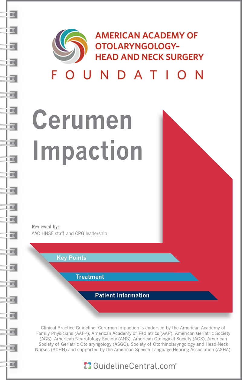
Cerumen Impaction
- 14 pages
- Spiral Bound
- 80# Aqueous Coating
- 4.25" x 7.25"
- Ships in 5 – 10 business days
- Key Points
- Images
- Anatomy
- Otoscopic views
- Tables
- Summary of Guideline Key Action Statements
- Options to Help Reduce Earwax
- Topical Preparations
- Proper care of Hearing aid(s)
- Shared Decision Grid For Patients and Caregivers For Cerumen Management
- Management Algorithm
- Patient Information
- Frequently Asked Questions
- Dos and Don’ts of Cerumen (Earwax)
- For purchases under 100 in quantity, we suggest placing the order directly through the website.
- We offer group/institutional licenses for multi-user accounts (discount amount varies depending on the number of users).
- We are proud to offer special discounts to medical schools, training programs, students and more.
- We offer bulk purchase discounts based on number of copies and number of titles.
Contact Us for more details
Description
This resource is for informational purposes only, intended as a quick-reference tool based on the cited source guideline(s), and should not be used as a substitute for the independent professional judgment of healthcare providers. Practice guidelines are unable to account for every individual variation among patients or take the place of clinician judgment, and the ultimate decision concerning the propriety of any course of conduct must be made by healthcare providers after consideration of each individual patient situation. Guideline Central does not endorse any specific guideline(s) or guideline recommendations and has not independently verified the accuracy hereof. Any use of this resource or any other Guideline Central resources is strictly voluntary.
You can also find this product included in this bundle!

AAO-HNS Guidelines Bundle
Last Updated: Feb 27, 2024- Acute Otitis Externa
- Adult Sinusitis
- Allergic Rhinitis
- Bell’s Palsy
- Benign Paroxysmal Positional Vertigo
- Cerumen Impaction
- Evaluation of the Neck Mass in Adults
- Hoarseness (Dysphonia)
- Immunotherapy for Inhalant Allergy
- Improving Nasal Form and Function after Rhinoplasty
- Improving Voice Outcomes After Thyroid Surgery
- Ménière’s Disease
- Nosebleed (Epistaxis)
- Opioid Prescribing for Analgesia After Common Otolaryngology Operations
- Otitis Media with Effusion
- Sudden Hearing Loss
- Tinnitus
- Tonsillectomy in Children
- Tympanostomy Tubes in Children
Related Guidelines
Immunotherapy for Inhalant Allergy
Tympanostomy Tubes in Children
Opioid Prescribing for Analgesia After Common Otolaryngology Operations
Ménière’s Disease
Nosebleed (Epistaxis)
Sudden Hearing Loss
Tonsillectomy in Children
Hoarseness (Dysphonia)
Evaluation of the Neck Mass in Adults
Benign Paroxysmal Positional Vertigo
Improving Nasal Form and Function after Rhinoplasty
Otitis Media with Effusion
Adult Sinusitis
Allergic Rhinitis
Improving Voice Outcomes After Thyroid Surgery
Tinnitus
Acute Otitis Externa
