
Peripheral Artery Disease Lower Extremity
- 28 pages
- Spiral Bound
- 80# Aqueous Coating
- 4.25" x 7.25"
- Ships in 5 – 10 business days
- Key Points
- Definitions
- History/Physical Examinations Suggestive of PAD
- Clinical Assessment for PAD
- Physiological Testing
- Alternative Diagnoses for Leg Pain or Claudication
- Alternative Diagnoses for Non-Healing Wounds
- Diagnostic Testing
- Imaging and Anatomical Assessment
- Antiplatelet Agents
- Statin Agents
- Antihypertensive Agents
- Smoking Cessation
- Glycemic Control
- Oral Anticoagulation
- Chelation Therapy
- Homocysteine Lowering
- Influenza vaccination
- Structured Exercise Therapy
- Minimizing Tissue Loss
- Revascularization for claudication
- Endovascular Revascularization for claudication
- Interdisciplinary Care Team for PAD
- For purchases under 100 in quantity, we suggest placing the order directly through the website.
- We offer group/institutional licenses for multi-user accounts (discount amount varies depending on the number of users).
- We are proud to offer special discounts to medical schools, training programs, students and more.
- We offer bulk purchase discounts based on number of copies and number of titles.
Contact Us for more details
Our mission is to build healthier lives, free of cardiovascular diseases and stroke. That single purpose drives all we do. AHA's Professional Membership is a made up of a robust group of cardiovascular professionals who participate in discovery and dissemination of science.
Description
This resource is for informational purposes only, intended as a quick-reference tool based on the cited source guideline(s), and should not be used as a substitute for the independent professional judgment of healthcare providers. Practice guidelines are unable to account for every individual variation among patients or take the place of clinician judgment, and the ultimate decision concerning the propriety of any course of conduct must be made by healthcare providers after consideration of each individual patient situation. Guideline Central does not endorse any specific guideline(s) or guideline recommendations and has not independently verified the accuracy hereof. Any use of this resource or any other Guideline Central resources is strictly voluntary.
You can also find this product included in these bundles!
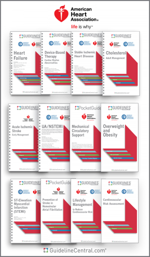
AHA Guidelines Bundle
Last Updated: Nov 30, 2023- Coronary Artery Revascularization
- CPR for Mechanical Circulatory Support
- Device-Based Therapy
- Diagnosis and Management of Aortic Disease
- Diagnosis and Treatment of Patients with Hypertrophic Cardiomyopathy
- Early Management of Patients With Acute Ischemic Stroke
- Evaluation and Diagnosis of Chest Pain
- Evaluation and Management of Patients with Bradycardia and Cardiac Conduction Delay
- Evaluation and Management of Patients With Syncope
- Heart Failure
- Management of Adults with Congenital Heart Disease
- Management of Blood Cholesterol
- Management of Patients with Atrial Fibrillation
- Management of Patients with Chronic Coronary Disease
- Management of Patients With Ventricular Arrythmias and the Prevention of Sudden Cardiac Death
- Mechanical Circulatory Support
- Mechanical Circulatory Support — Ambulatory and Community Patient Care
- Non-ST-Elevation Acute Coronary Syndromes
- Perioperative Cardiovascular Evaluation and Management of Patients Undergoing Noncardiac Surgery
- Peripheral Artery Disease Lower Extremity
- Prevention Of Stroke In Nonvalvular Atrial Fibrillation
- Prevention of Stroke In Women
- Prevention, Detection, Evaluation, and Management of High Blood Pressure in Adults
- Primary Prevention of Cardiovascular Disease
- Primary Stroke Prevention
- ST-Elevation Myocardial Infarction (STEMI)
- Stable Ischemic Heart Disease
- Supraventricular Tachycardia
- Valvular Heart Disease
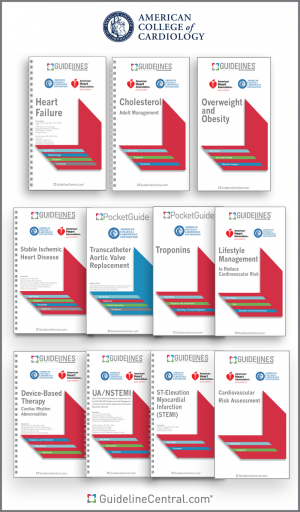
ACC Guidelines Bundle
Last Updated: Nov 30, 2023- Coronary Artery Revascularization
- Device-Based Therapy
- Diagnosis and Management of Aortic Disease
- Diagnosis and Treatment of Patients with Hypertrophic Cardiomyopathy
- Evaluation and Diagnosis of Chest Pain
- Evaluation and Management of Patients with Bradycardia and Cardiac Conduction Delay
- Evaluation and Management of Patients With Syncope
- Heart Failure
- Management of Adults with Congenital Heart Disease
- Management of Blood Cholesterol
- Management of Patients with Atrial Fibrillation
- Management of Patients with Chronic Coronary Disease
- Management of Patients With Ventricular Arrythmias and the Prevention of Sudden Cardiac Death
- Non-ST-Elevation Acute Coronary Syndromes
- Perioperative Cardiovascular Evaluation and Management of Patients Undergoing Noncardiac Surgery
- Peripheral Artery Disease Lower Extremity
- Prevention, Detection, Evaluation, and Management of High Blood Pressure in Adults
- Primary Prevention of Cardiovascular Disease
- ST-Elevation Myocardial Infarction (STEMI)
- Stable Ischemic Heart Disease
- Supraventricular Tachycardia
- Valvular Heart Disease
Related Guidelines
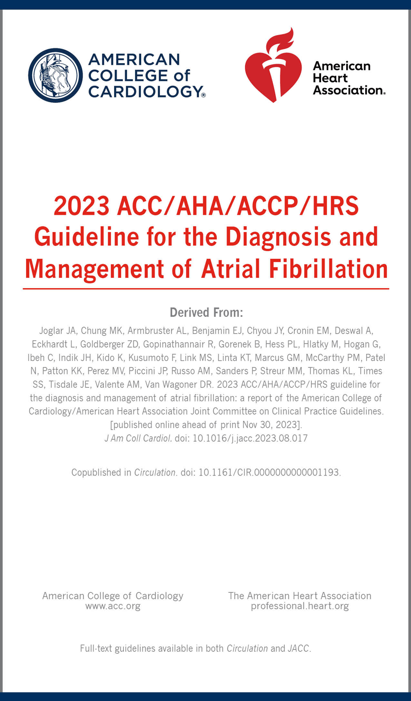
Management of Patients with Atrial Fibrillation
Last Updated: Nov 30, 2023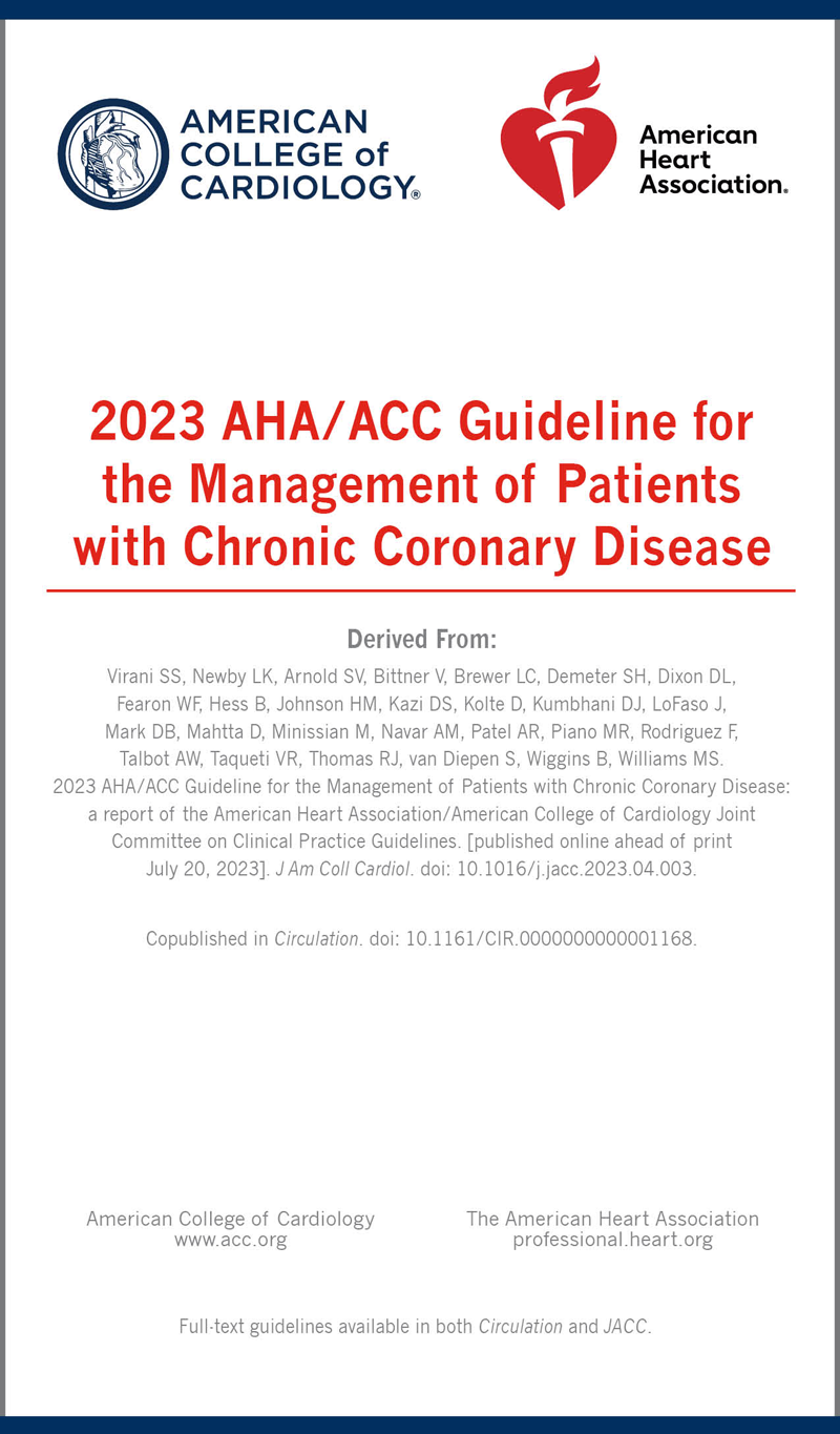
Management of Patients with Chronic Coronary Disease
Last Updated: Jul 20, 2023
Diagnosis and Management of Aortic Disease
Last Updated: Nov 2, 2022
Heart Failure
Last Updated: Mar 31, 2022
Coronary Artery Revascularization
Last Updated: Dec 9, 2021
Evaluation and Diagnosis of Chest Pain
Last Updated: Oct 28, 2021
Management of Adults with Congenital Heart Disease
Last Updated: Apr 20, 2021
Evaluation and Management of Patients With Syncope
Last Updated: Apr 18, 2021
Evaluation and Management of Patients with Bradycardia and Cardiac Conduction Delay
Last Updated: Mar 16, 2021
Valvular Heart Disease
Last Updated: Dec 20, 2020
Diagnosis and Treatment of Patients with Hypertrophic Cardiomyopathy
Last Updated: Nov 19, 2020
Early Management of Patients With Acute Ischemic Stroke
Last Updated: Mar 4, 2020
Primary Prevention of Cardiovascular Disease
Last Updated: Oct 15, 2019
Management of Blood Cholesterol
Last Updated: Feb 11, 2019
Prevention, Detection, Evaluation, and Management of High Blood Pressure in Adults
Last Updated: Dec 20, 2018
Management of Patients With Ventricular Arrythmias and the Prevention of Sudden Cardiac Death
Last Updated: Dec 18, 2018
CPR for Mechanical Circulatory Support
Last Updated: Oct 30, 2017
Mechanical Circulatory Support — Ambulatory and Community Patient Care
Last Updated: Oct 30, 2017
Supraventricular Tachycardia
Last Updated: Nov 11, 2015
Non-ST-Elevation Acute Coronary Syndromes
Last Updated: Feb 11, 2015
Primary Stroke Prevention
Last Updated: Feb 10, 2015
Mechanical Circulatory Support
Last Updated: Jan 6, 2015
Perioperative Cardiovascular Evaluation and Management of Patients Undergoing Noncardiac Surgery
Last Updated: Jan 6, 2015
Prevention of Stroke In Women
Last Updated: Jan 6, 2015
Stable Ischemic Heart Disease
Last Updated: Jan 6, 2015
ST-Elevation Myocardial Infarction (STEMI)
Last Updated: Jan 5, 2015
Prevention Of Stroke In Nonvalvular Atrial Fibrillation
Last Updated: Jan 5, 2015